Anatomy and Working of the Eye and Refractive Errors
The eye enables sight – one of the 5 primary senses in the human body. Hence, protecting your eyesight is extremely important. Consuming nutritious meals and regularly visiting your ophthalmologist ensures that you maintain eye health and retain your sight.
Understanding the anatomy of the eye helps you maintain its health. It also allows you to identify any eye problems you may have. Alongside an expert ophthalmologist like Dr Anisha Gupta, you can easily and effectively treat any eye problems you may have with guaranteed results.

The Anatomy of the Eye
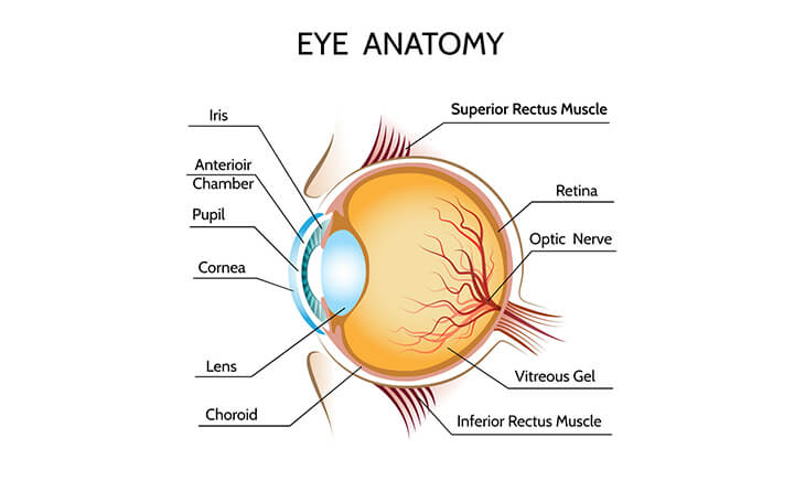
From the front to the back, 12 parts optimise eye function. Let us explain these to you in order:
These essential structures ensure that your eye continues to function perfectly. If any of these are affected, the vision is affected.
The Working of the Eye
Understanding the eye’s working is very similar to operating a camera. Much like the camera’s shutter, the eye’s aperture, the pupil, opens or closes depending on the amount of light. In a camera, this light exposes the film at the back of the camera. For the eye – the iris and pupil work together to allow light to the back of the eye.
Our eyesight depends on the transfer of light through the front of the eye (the cornea and lens) to the retina. Together, the cornea and lens focus the light rays onto the retina. The cells in the macula then absorb and convert the light into electrochemical impulses. These are transferred to the brain via the optic nerve. In the process, the optic nerve has to turn the images right side up since the retina perceives the images upside down.
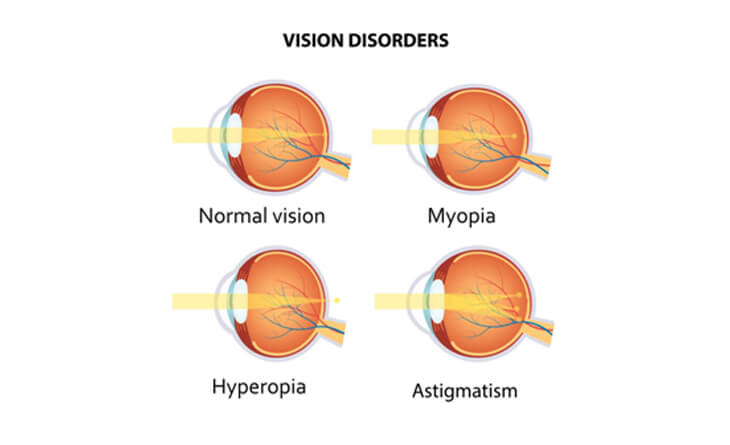
Refractive errors are conditions in which the eye is largely normal in structure, but light does not get focused at the retina. Hence, we need glasses or contact lenses to make the light focus at the right place (retina).
Refractive Errors and Their Types
If you have blurry vision, you may be experiencing a refractive error. A refractive error is when your eye cannot properly bend light and focus it on the retina. It may happen because your eyeball is too long or short, your cornea is improperly shaped, or your lens has aged and become rigid and clouded.
Its symptoms include:
If you experience any of these, please contact an ophthalmologist immediately and seek your course of action. If left untreated, refractive errors could cause further complications in your eye.
There are also different types of refractive errors. The 4 most common are:

Refractive errors can be corrected easily and safely with glasses, contact lenses or surgery. With an expert like Dr Anisha Gupta, you get excellent results with accurate diagnoses and effective treatment paths.
Dr Gupta uses the latest techniques and devices for excellent outcomes that minimise downtime and promote a comfortable, speedy recovery!
Contact us today and check your eye health with our expert ophthalmologist!
